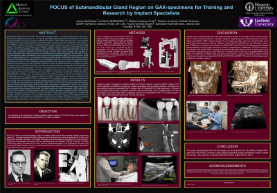
Purpose of the Study: POCUS is safe, non-invasive imaging useful in multiple dental arenas from general dentistry diagnosing caries etc…to implant specialists aiding diagnosis and treatment from tissue acquisition(soft & bony) and analysis for effective outcomes. Oral health clinicians have demonstrated a propensity for improving techniques and technologies optimizing care for individuals and communities. POCUS continues to evolve customizing training and research on neck, oral and maxillofacial regions. The objective of this study was to identify a lifelike cadaver medium for POCUS training and research of the submandibular gland/fossa region(SGFR) by implant specialists.
Methods: Literature search was conducted regarding POCUS training of SGFR from donor- cadavers with contrast by implant specialists. GE-Vscan air wireless handheld ultrasound probes delivered POCUS. SGFR imaging was acquired(graded acceptable or poor concerning submandibular gland, mylohyoid muscle, facial artery, vessels/nerves) from a sonographer with GAX-specimens(n=19:38sides) and BriteVu contrast compared to formalin-fixed cadavers(F-FC),n=16:32sides. CBCT/CT scans were conducted for further assessment of POCUS with subsequent dissection.
Results: Literature search revealed no known studies of POCUS training acquiring SGFR images from donor-cadavers with/without contrast by implant specialists during surgical courses. GE-Vscan air successfully acquired acceptable SGFR images from 37/38sides(97%) GAX-specimens. 1/38 was poor probably due to being a cephalic specimen versus entire body. Only 2/32sides(6%)F-FC were acceptable, 30/32 were poor due to collapsed vessels and dehydration from F-FC embalming process. CBCT and dissection confirmed POCUS structures.
Conclusion: In 2022, it was reported more than 150 million people in the US are missing at least 1-tooth and just over 1 million received between 3-5million implants. Regarding bone measurements of submandibular fossa(SM-f), CBCT is gold standard for pre-implant workup, however, its significantly more costly, time-consuming and delivers radiation versus POCUS. POCUS can be alternative evaluation of SM-f for pre-implant workup/treatment. Both CBCT and POCUS can aid in preventing lingual plate perforation during surgery resulting in serious complications. Additionally, POCUS can evaluate infection and lymph nodes. Imaging of submandibular gland helps prevent cortical lingual plate perforation protecting neurovascular structures. No other known study has investigated SGFR using CBCT and POCUS on lifelike GAX-specimens with BriteVu contrast. Edentulous implant patient cohorts need research assessing the SGFR with POCUS and CBCT. This study methodology would enable this valuable research. This study demonstrated that POCUS images can be acquired from the SGFR of lifelike GAX-specimens with BriteVu contrast to train implant specialists during conventional implant surgery workshops. This would be a valuable alternative and/or supplement to CBCT.
Articles: Akol Gorgun E, Caglayan F. Evaluation of submandibular gland and submandibular fossa: a combined cone beam computed tomography and ultrasound study. Eur Oral Res. 2023;57(3):128-132. doi:10.26650/eor.20231109135. PMID: 37929223; PMCID: PMC10622153.
Alghamdi AS. Pain sensation and postsurgical complications in posterior mandibular implant placement using ridge mapping, panoramic radiography, and infiltration anesthesia. ISRN Dent. 2013;2013:134210. doi:10.1155/2013/134210.
Çağlayan F, Ocak A, Sümbüllü MA. Supramandibular lymph nodes in dental patients by ultrasonography. Nobel Med. 2019;15(1):5-10.
Damstra J, Fourie Z, Huddleston Slater JJ, Ren Y. Accuracy of linear measurements from cone-beam computed tomography–derived surface models of different voxel sizes. Am J Orthod Dentofacial Orthop. 2010;137(1):16.e1-16.e6. doi:10.1016/j.ajodo.2008.09.025.
Clinical Applications of Ultrasound Imaging in Dentistry: A Comprehensive Literature Review. Imaging Sci Dent. 2024. Accessed October 8, 2025. https://www.sciencedirect.com/science/article/pii/S2772559624000099
Overview of Ultrasound in Dentistry for Advancing Research Methodology and Patient Care Quality with Emphasis on Periodontal/Peri-implant Applications. Clin Oral Investig. 2023. Accessed October 8, 2025. https://www.sciencedirect.com/science/article/pii/S0939388923000053
Gandage SG, Kachewar SG. An imaging panorama of salivary gland lesions as seen on high-resolution ultrasound. J Clin Diagn Res. 2014;8(3):RC01-RC13. doi:10.7860/JCDR/2014/7409.4153.
Isaacson TJ. Sublingual hematoma formation during immediate placement of mandibular endosseous implants. J Am Dent Assoc. 2004;135(2):168-172. doi:10.14219/jada.archive.2004.0161.
Kobayashi K, Shimoda S, Nakagawa Y, Yamamoto A. Accuracy in measurement of distance using limited cone-beam computerized tomography. Int J Oral Maxillofac Implants. 2004;19(2):228-231.
Niamtu J III. Near-fatal airway obstruction after routine implant placement. Oral Surg Oral Med Oral Pathol Oral Radiol Endod. 2001;92(6):597-600. doi:10.1067/moe.2001.119127.
Philipsen HP, Takata T, Reichart PA, Sato S, Suei Y. Lingual and buccal mandibular bone depressions: a review based on 583 cases from a worldwide literature survey, including 69 new cases from Japan. Dentomaxillofac Radiol. 2002;31(5):281-290. doi:10.1038/sj.dmfr.4600713.
Stratemann SA, Huang JC, Maki K, Miller AJ, Hatcher DC. Comparison of cone beam computed tomography imaging with physical measures. Dentomaxillofac Radiol. 2008;37(2):80-93. doi:10.1259/dmfr/13470511.
Zengel P, Schrötzlmair F, Reichel C, Paprottka P, Clevert DA. Sonography: the leading diagnostic tool for diseases of the salivary glands. Semin Ultrasound CT MR. 2013;34(3):196-203. doi:10.1053/j.sult.2012.12.002.
Books: Standring S, ed. Gray’s Anatomy: The Anatomical Basis of Clinical Practice. 42nd ed. Philadelphia, PA: Elsevier; 2020.