Clinical Associate Professor, Division Director of Clinical Implantology East Carolina School of Dental Medicine Greenville, North Carolina, United States
Purpose of the Study: Recent developments in digital and implant dentistry, have allowed the clinicians to utilize computer-aided design and computer-aided manufacturing (CAD/CAM) systems for a fully digital protocol for both surgical and restorative phases of implant therapy and thus increase implant success. As part of this treatment process, the use of customized healing abutments has been implemented with the purpose of contouring the soft tissues and creating a natural emergence profile that would allow for an esthetic and functional implant restoration. The purpose of these case series is to validate a digital protocol to fabricate customized CAD/CAM healing abutments that will improve the predictability of the final restoration and delivery, as well as soft tissue contours, bone stability, peri-implant health, and patient satisfaction.
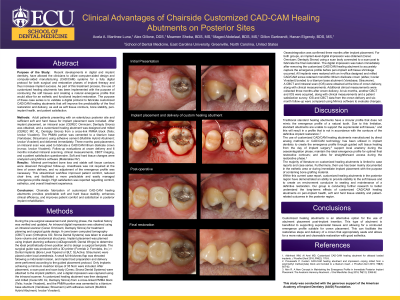
Methods: Adult patients presenting with an edentulous posterior site and sufficient soft and hard tissue for implant placement were included. After implant placement, an intraoral scan (CEREC Omnicam; Dentsply Sirona) was obtained, and a customized healing abutment was designed and milled (CEREC MC XL; Dentsply Sirona) from a cross-link PMMA block (Telio; Ivoclar Vivadent). The PMMA portion was cemented to a titanium base (Variobase; Straumann) using adhesive cement (Multilink Hybrid Abutment; Ivoclar Vivadent) and delivered immediately. Three months post-placement, an intraoral scan was used to fabricate a CAD/CAM lithium disilicate crown (e.max; Ivoclar Vivadent). Follow-up evaluations at crown delivery and 6 months included intraoral scanning, clinical measurements, CBCT imaging, and a patient satisfaction questionnaire. Soft and hard tissue changes were analyzed using Mimics software (Materialise NV).
Results: Minimal peri-implant bone loss and stable soft tissue contours were observed throughout follow-up. Anesthesia was not required at the time of crown delivery, and no adjustment of the emergence profile was necessary. This streamlined workflow improved patient comfort, reduced chair time, and facilitated a more predictable and easily managed emergence profile design. High satisfaction was reported regarding comfort, esthetics, and overall treatment experience.
Conclusion: Chairside fabrication of customized CAD-CAM healing abutments provides predictable soft and hard tissue stability, enhances clinical efficiency, and improves patient comfort and satisfaction in posterior implant rehabilitation.
Articles: Alshhrani WM, Al Amri MD. Customized CAD-CAM healing abutment for delayed loaded implants. J Prosthet Dent. 2016;116(2):176-179. Proussaefs P. Custom CAD-CAM healing abutment and impression coping milled from a poly(methyl methacrylate) block and bonded to a titanium insert. J Prosthet Dent. 2016;116(5):657-662. Akin R. A new concept in maintaining the emergence profile in immediate posterior implant placement: The Anatomic Harmony Abutment. J Oral Maxillofac Surg. 2016;74(12):2385-2392.