(P12) Masseter and Medial Pterygoid Muscles Are Actually a Functional Unit Connected by a Tendon with Minimal Osseous Insertion Termed the Zygopterygoid Muscle
Purpose of the Study: Masseter is recognized as the strongest muscle in the body regarding mass to force ratio and exercised daily via mastication and speech maintaining impressive power, yet no distal tubercle has developed, and arguably only a minimal ridge may exist at best. Imagine the ramus and angle of mandible transparent, one would essentially see a mirror image of 2 muscle bellies with fibers angled similarly (masseter and medial pterygoid) connected via a musculotendinous sling with minimal osseous attachment. Benninger developed a dissection technique which includes light manual finger-tip dissection separating distal masseter and medial pterygoid away from the inferior mandible border and ramus region revealing musculotendinous unit. Two muscle bellies on either side of the ramus (lateral/medial orientation) with similar morphology including fiber direction connected via musculotendinous sling warrants term consideration (Zygopterygoid muscle) describing 2 bellies as demonstrated by digastric and omohyoid muscles. The study objective is to provide evidence for naming the masseter and medial pterygoid as Zygopterygoid muscle to be recognized and taught as a dual muscle unit with musculotendinous sling as formal terminology from dissection, surgery and radiology.
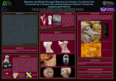
Methods: Literature search was conducted on contemporary anatomy texts/atlases and surgical texts/literature regarding pterygomasseter sling or Zygopterygoid muscle with lateral/medial heads. Benninger dissection technique(BDT) includes controlled finger-tip separation of distal masseter-medial pterygoid muscles from inferior mandibular angle-border region with GAX-specimens(n=19:38-sides) with BriteVu-contrast for CT/MRI, fresh-frozen- cadavers(FreshFC)n=18:36sides and formalin-fixed-cadavers(F-FC)n=445:879sides,(11-sides were cut), overall total n=482:953sides.
Results: Literature search revealed no contemporary anatomy texts/atlases describing or illustrating pterygomasseter sling or Zygopterygoid muscle with lateral/medial heads. Anatomy websites mention pterygomasseter sling. Surgical texts/procedures consistently described pterygomasseter-sling. GAX-specimens, FreshFC, F-FC received BDT to 953- sides revealing tendinous connection or sling. The tendon connecting 2 muscles demonstrated length variability and heterogenicity of muscle fibers within the tendon. Interestingly consistent small tendon attachments (2-3) were noted in the lower one-third of ramus body, demonstrating an element of tacking the muscle down to perhaps prevent anterior-poster sliding.
Conclusion: Cross-sectional imaging has consistently demonstrated tendinous sling connecting masseter-medial-pterygoid muscles. Eschmarc described the pterygo-masseteric
sling in a surgical technique. Existing anatomical patterns describe dual muscle belly structures with intermediate tendon at the transition from thorax to neck(omohyoid), transition from neck to mandible(digastric), transition from mandible to face/skull(Zygopterygoid or masseter-medial pterygoid). As clinician-researcher-educators, lets improve communication, etc… This study revealed distal masseter-medial-pterygoid anatomy was connected by musculotendinous sling confirmed by dissection, imaging and surgery. suggesting formal teaching of the Zygopterygoid muscle.
Articles: Dmetrichuk JM, McLachlan SM, Laitman JT. Clinical importance of morphology and nomenclature of distal attachment of temporalis tendon. J Oral Maxillofac Surg. 2011;69(11):2896-2901. doi:10.1016/j.joms.2011.01.024
Books: Standring S, ed. Gray’s Anatomy: The Anatomical Basis of Clinical Practice. 42nd ed. Elsevier; 2020.