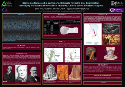
(P09) Sternocleidomastoid Is an Important Muscle for Extra Oral Examination Identifying Variations Before Dental Implants, Central Lines and Neck Surgery
Pre-Dental Student Center for Family and Implant Dentistry Gresham, Oregon, United States
Purpose of the Study: This study aimed to investigate the anatomical variations of the sternocleidomastoid (SCM) muscle in formalin-fixed cadavers, fresh-frozen cadavers, and GAX-prepared specimens. The SCM plays a fundamental role in extraoral examinations and serves as a critical landmark in numerous clinical procedures, including dental implant placement, temporomandibular joint assessment, central line insertions, and neck surgeries. Previous research indicates a 10–30% incidence of SCM variations, yet these findings are inconsistently reflected in contemporary anatomy texts and atlases. By documenting the incidence and morphology of SCM variants, this study seeks to bridge the gap between research literature and clinical practice, improve anatomical education, and increase awareness of the clinical importance of SCM variations to minimize morbidity and optimize patient outcomes.
Methods: A literature review of sternocleidomastoid (SCM) anatomy was conducted using atlases, textbooks, and online resources to identify reported attachment variations. Anatomical dissections were performed on 45 formalin-fixed cadaver donors (90 sides; 27 male, 18 female) and 7 GAX-prepared specimens (14 sides; 4 male, 3 female). Each SCM was examined for superior and inferior attachment points, compared with documented references, and classified as normal or variant. The incidence of SCM variations was calculated, and findings were analyzed against published anatomical descriptions to assess discrepancies between research data and contemporary educational sources.
Results: The literature review revealed inconsistent descriptions of the sternocleidomastoid (SCM), particularly regarding superior attachment points, and minimal recognition of inferior attachment variations. Dissection of 104 SCM muscles (90 from formalin-fixed cadavers, 14 from GAX-prepared specimens) identified 11 variant sides (11%). Variations were observed in 7 of 52 donors (13%), with 4 bilateral cases (accounting for 73% of all variants, 8 sides) and 3 unilateral cases (27%, 3 sides). These findings suggest that SCM variations may be more prevalent than indicated in standard anatomical texts, which often lack detailed clinical correlation. The results support research literature estimating SCM variation incidence between 10–30% and underscore the importance of recognizing these variants in both anatomical education and clinical practice.
Conclusion: This study highlights a disconnect between contemporary anatomy texts and research literature regarding sternocleidomastoid (SCM) morphology and its clinical relevance. Variations of the SCM were identified in 13% of cadaver donors, supporting published reports of a 10–30% incidence. Given the SCM’s role as a critical landmark for dental implant placement, temporomandibular joint assessment, central line insertion, and neck surgery, awareness of its variations is essential to minimize procedural morbidity. Larger, more comprehensive studies are warranted to establish true incidence; however, increased attention to SCM anatomy in clinical education can enhance diagnostic accuracy, surgical safety, and patient outcomes.
Articles: Sinnatamby CS. Last’s Anatomy: Regional and Applied. 11th ed. Edinburgh, Scotland: Churchill Livingstone/Elsevier; 2006. Snell RS. Clinical Anatomy by Regions. 7th ed. Philadelphia, PA: Lippincott Williams & Wilkins; 2004.
Books: Drake RL, Vogl AW, Mitchell AWM. Gray’s Anatomy for Students. 2nd ed. Philadelphia, PA: Churchill Livingstone/Elsevier; 2010. Moore KL, Dalley AF, Agur AMR. Clinically Oriented Anatomy. 7th ed. Philadelphia, PA: Wolters Kluwer/Lippincott Williams & Wilkins; 2014. Netter FH. Atlas of Human Anatomy. 7th ed. Philadelphia, PA: Elsevier; 2019. Ranganathan TS. A Textbook of Human Anatomy. 6th ed. New Delhi, India: S. Chand & Company Ltd, 2008.